
Case Studies: SPECT-CT in Practice
A small selection of representative cases demonstrating how HealthyPaws works with referring veterinarians to clarify complex diagnostic questions.
The following cases are intended to illustrate how SPECT-CT can add value in complex or inconclusive presentations. Each example reflects a collaborative diagnostic process between Healthy Paws and the referring veterinarian.
- Orthopaedics
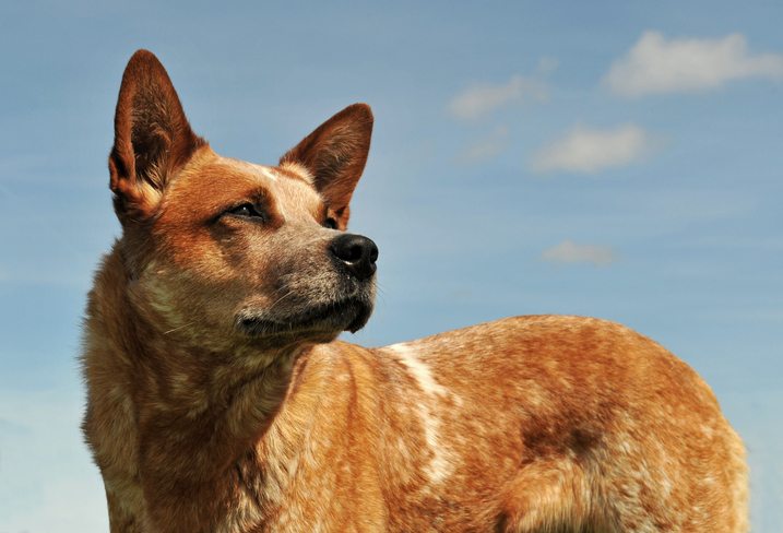
Persistent Hock Pain in a Racing Greyhound
Clinical Challenge:
3-year-old racing Greyhound with a previously diagnosed stress fracture of the right hock (tarsus), identified on radiographs following an acute onset lameness during training. The fracture was managed conservatively with rest and rehabilitation, and follow-up imaging indicated appropriate healing. The dog was cleared to return to racing.

Why Conventional Imaging Was Inconclusive:
Standard X-rays confirmed healing of the original stress fracture and showed no clear evidence of catastrophic injury. Subtle bone stress reactions or ongoing metabolic bone activity are often not visible on routine radiographs once structural healing appears complete. As a result, there was uncertainty as to whether the lameness was behavioural, soft tissue–related, or due to unresolved bone pain.
What SPECT-CT Revealed:
Functional imaging demonstrated persistent focal increased uptake within the right hock, consistent with ongoing bone stress and active remodelling at the previous fracture site. Additional low-grade uptake was also noted in adjacent tarsal structures, indicating that the joint had not fully returned to a normal metabolic state despite radiographic healing. This confirmed that the dog was still experiencing active pathology and likely discomfort during high-intensity performance.
Outcome & Treatment Planning:
The dog was formally retired from racing, transitioned into a pet home, and managed with supportive joint care and controlled exercise. In the lower-demand environment, the Greyhound has remained comfortable and has successfully adapted to life as a companion animal, demonstrating the value of advanced imaging in supporting evidence-based and welfare-focused decision-making.This confirmed that the dog was still experiencing active pathology and likely discomfort during high-intensity performance.
- Oncology
Dog Cancer Case Study: Tracking Sarcoma & Hip Pain in a 10-Year-Old Kelpie
Clinical Challenge:
A much-loved 10-year-old Kelpie × Red Heeler had recently undergone surgery to remove a sarcoma from the right fore elbow. While the mass had been excised, the referring veterinary team remained concerned that microscopic cancer cells could still be present. On top of this, the dog had lived with hip dysplasia since 18 months of age, and mobility had been gradually declining. The question was - how much of the discomfort was coming from the hips, and was there still active cancer at the surgical site?
Why Standard Imaging Wasn’t Enough
After surgery, the elbow region showed normal post-operative changes - swelling, healing tissue and scar formation. On conventional imaging, it was difficult to confidently determine whether any tumour cells remained active. Radiographs confirmed known hip dysplasia, but they could not clearly show which hip was contributing most to pain, or whether there was active inflammation driving the mobility decline. When anatomy alone cannot tell the full story, functional imaging becomes critical.
What SPECT-CT Revealed:
SPECT-CT provided both metabolic activity and anatomical localisation.
The scan identified:
• Ongoing metabolic activity in the right fore elbow, consistent with residual sarcoma cells
• More significant degenerative change and active inflammation in the right hip compared to the left
Rather than guessing, the team could now see exactly where disease was still active.
What Happened Next
With confirmation of residual tumour activity, electrochemotherapy was initiated to directly target remaining sarcoma cells. The hip dysplasia management plan was also refined, focusing on the right hip as the primary pain source and optimising long-term comfort and mobility support.
Why This Matters
In complex cases involving both cancer surveillance and chronic orthopaedic disease, SPECT-CT helps answer one simple but critical question: Where is the disease still active?
Each case reflects a collaborative interpretation process between Healthy Paws and the referring veterinarian, supporting shared decision-making and improved diagnostic confidence.
Have a Case You’d Like to Discuss?
If you’re managing a complex or unclear case, the Healthy Paws team is available to discuss whether SPECT-CT may be helpful.